What is an Eosinophilic Granuloma?
Eosinophilic Granuloma is a type of damage that occurs on bones (lesions) due to an overgrowth of immune cells called Langerhans cells. These cells can malfunction and develop into bone tumors on different bones and different areas on those bones. If multiple lesions occur, then there is a risk of organs being involved
Biological Behavior
- • This disorder typically acts benign but can cause damage to bones and eventually organs.
- • Recurrence can potentially happen, but is uncommon.
Causes
- • Some studies link this condition to a mutation in the BRAF V600E gene and/or MAP2K1 mutations.
Signs and Symptoms
- • Pain and swelling in the region of the affected area
- • Elevated levels of white blood cells (eosinophilia)
- • Temporal bone disease
- • Fever
Common Bones Involved
- • Skull
- • Pelvis
- • Humerus
- • Flat Bones (front of face)
Who is usually affected?
- • 85% of case occur anywhere from birth to 30 years old.
- • 60% of cases occur from birth to 10 years old.
- • Men are more commonly affected.
- • Rare; 4 to 5 case per million a year in children and 1 to 2 cases per million a year in adults.
Diagnosis
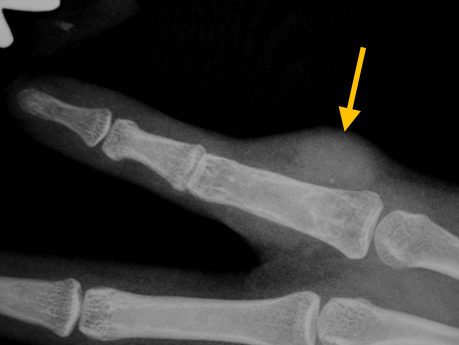
- • The work-up often consists of a physical examination, X-rays, CT scans, MRI, and sometimes bone scans are required.
- • The diagnosis is often confirmed with a biopsy, which means taking a sample of tumor and having it analyzed under a microscope by a pathologist.
Risk to your limbs
Eosinophilic Granulomas are benign tumors that lead to bone destruction and potential organ involvement.
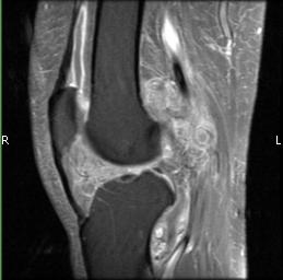
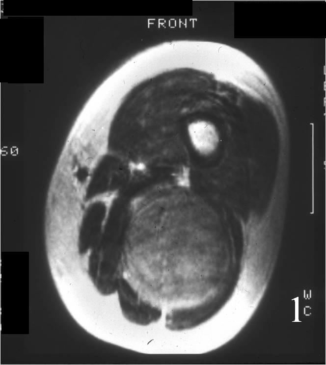
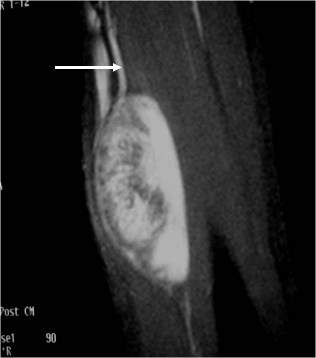
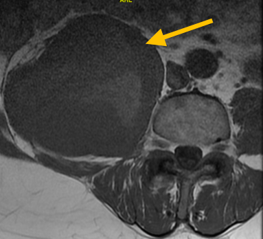
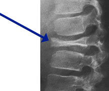
Radiographic imaging is used to help form a diagnosis. These include X-Ray, MRI, CT and Bone Scans.
An example of an Eosinophilic Granuloma MRI is shown.
Treatment of Eosinophilic Granuloma
The majority of patients are cured by scooping the tumor out of the bone (curettage) and bone grafting or injecting a steroid within the tumor. Low dose radiation may also be used for damage to the bone (lesion) that is unaccessible.
Intralesional Curettage
Intralesional Curettage means to scoop the tumor out using a spoon-like tool called a curette. This is a surgery that aims to remove the mass and restore the bone so that the patient can get back to normal function. The eosinophilic granuloma is identified within the bone and scooped, or curetted, out. The cavity is then shaved down with a Midas Rex Drill, which is similar to a dental drill. This drill removes more tumor cells.
Radiation
Radiation therapy is a localized treatment that utilizes high-energy particles or waves to kill cancerous cells. Because radiation therapy is a localized treatment, it only affects the area in which it is set to target and therefore eliminates the risks of damaging healthy cells throughout the body. Not only is it used to treat cancer, but it can also decrease the chances of the cancer from recurring. Lastly, radiation may be used in conjunction with other treatments, such as surgery or chemotherapy.
Bone Grafting and Fixation
The empty bone cavity is usually filled with bone graft or bone cement. Bone can be donated (allograft) or taken from the patient themselves (autograft). Fixation devices, such as a plate and screws, may be used in specific situations to prevent postoperative fracture.