What is Infantile Fibromatosis?
Infantile fibromatosis is a rare condition that arises from mesenchymal cells and involves the growth of a singular or numerous benign (non-cancerous) tumors. This condition is also referred to as infantile myofibromatosis or congenital fibromatosis. Tumors may be present at or shortly after birth, however a majority of all diagnoses occur before the age of two. They appear as firm, round nodules commonly located in the head, neck, trunk, and upper and lower extremities. While they are considered locally aggressive tumors, infantile fibromatosis does not metastasize, or spread to other body parts.
Who is usually affected?
- • Children under the age of 5, with a majority of cases diagnosed in children 2 years old or younger.
- • Boys are more commonly affected than girls with a ration of 2:1-3:1.
Common Sites Involved
- • Shoulder girdle
- • Muscles and fascia of the head/neck and thigh
- • Involvement of the long bones may be associated with tumors of the leg
Causes
- • The cause of infantile fibromatosis is unknown.
Signs and Symptoms
- • Signs and symptoms include a firm, growing mass.
Biological Behavior
- • Localized: most common, more commonly seen in males, 7-10% recurrence after excision, great prognosis.
- • Multicentric without visceral involvement: presents with multiple nodules.
- • Generalized form: multiple nodules with visceral involvement (lungs and GI tract), poorer prognosis.
Diagnosis
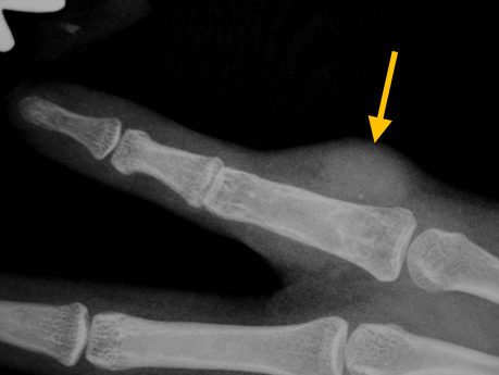
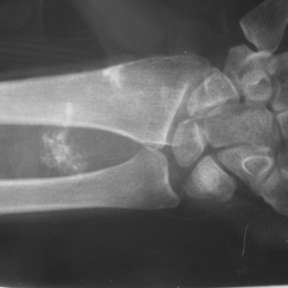
- • The work-up for Infantile Fibromatosis often consists of a physical examination, X-ray, MRI, CT, and bone scans. The diagnosis is often confirmed with a biopsy, which samples the tumor for further analysis. Differential diagnoses include hemangiomas, infantile fibrosarcoma, and other soft-tissue tumors.
Risk to your limbs
Infantile fibromatosis is a rare condition in which one or many non-cancerous tumors growth. These masses are considered locally aggressive tumors, however infantile fibromatosis does not metastasize, or spread to other body parts. On rare occasions, the tumor may grow back.
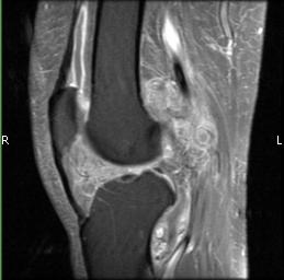
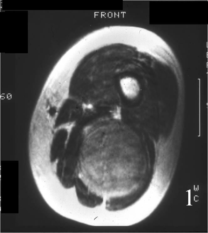
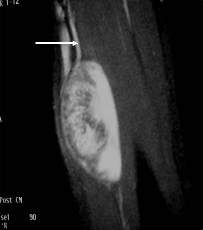
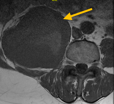
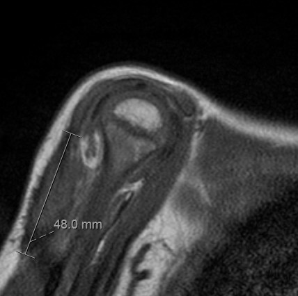
Radiographic imaging is used to help form a diagnosis of infantile fibromatosis. These include X-Ray, MRI, CT and Bone Scans.
An example of an MRI is shown.
Treatment of Infantile Fibromatosis
Treatment of infantile fibromatosis includes surgical excision of the tumor through wide or radical resections, both of which are limb-sparing surgeries. The tumor is removed as carefully and completely as possible without interfering with function or causing disfigurement to the affected area.
Surgery
Surgical treatment includes wide or radical resections to remove the complete tumor and additional margins. The removal of additional, surrounding margins ensures that the tumor is completely removed and decreases the chances of the tumor coming back.