What is a Pelvic Resections?
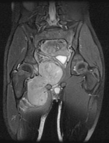
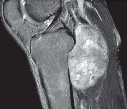
The pelvis is a common site for malignant and benign tumors to arise. The pelvis is composed of three parts; the ischium, illium, and pubis. The socket (hip joint) is called the acetabulum which forms the “cup.” Common primary sarcomas that can arise from the pelvis include osteosarcoma, Ewing’s Sarcoma, and chondrosarcoma. Soft tissue malignant tumors can sometimes involve the pelvis as well. Additionally, metastatic disease can commonly spread to the pelvis. Limb-sparing surgery can be performed for approximately 95% of tumors arising from the pelvis. In some instances the extremity cannot be saved and an amputation is performed.
Contraindications for saving the limb may include neurovascular invasion, infection, pathological fracture, extensive disease, contamination from a poorly performed biopsy, recurrent disease.
What’s involved in the technique?
Incision
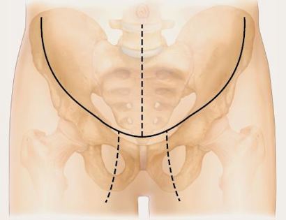
An incision is made that curves across the top of the femur bone.
Muscle Releases
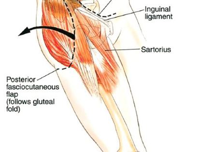
Developing surgical planes (margins that are tumor free) and separating muscles that can be preserved and leaving those in continuity with the tumor that should be removed, such as the gluteus muscles and iliac. This is based on preoperative MRI and intraoperative findings as well as the type of tumor.
Blood Vessel and Nerve Dissection
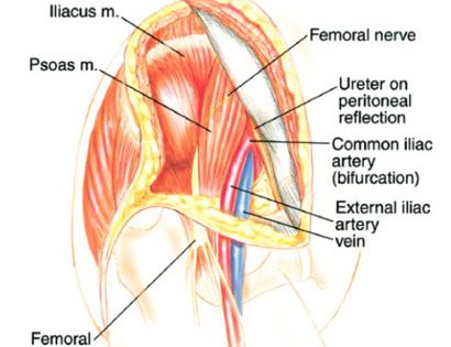
The sciatic nerve is identified and preserved as well as the femoral nerve. The nerves and blood vessels associated with the tumor or embedded in the tumor are ligated (closed off) and removed.
Tumor Removal and Reconstruction
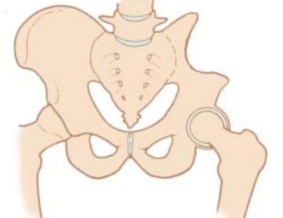
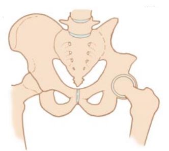
Removal of tumor and reconstruction. Once the tumor is removed the bone and joint are restored (reconstructed) if the tumor involved the “ball and socket” region. A “cup” may be utilized to replace the acetabulum if it was removed during surgery.
Soft Tissue Coverage
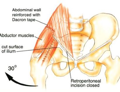
To reconstruct the hip after the tumor is removed, the gluteus medius muscle is attached to the abdomen as well as the adductors muscle. The sutures may be reinforced with surgical tape.
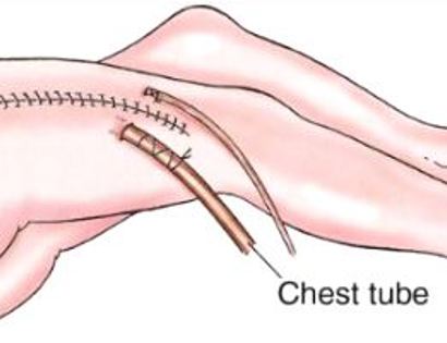
Closure
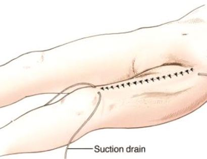
We then close your incision with sutures and cover the surgical site with bandages. Multiple large drains may also be used to drain the surgical site and prevent a seroma (buildup of fluid).
Capsule Closure
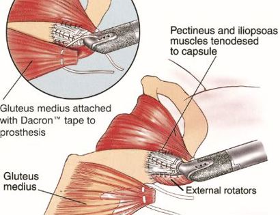
The gluteus medius muscle is attached to the prosthesis to ensure proper functioning post-surgically.
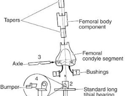
Distal Femur Components Schematic
Soft Tissue Coverage
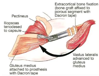
Multiple muscle rotation flaps are used to restore function and stability of the elbow as best as possible. The goal is to provide a stable hip so the extremity can function well. Soft-tissue reconstruction that involves rotating and reattaching the surrounding muscles, including the vastus lateralis, iliopsoas, and gluteus medius. Restoring the function of the surrounding muscles and hip joint is most important for achieving optimal functional outcomes and for protecting the prosthesis from infection.
Closure
We then close your incision with sutures and cover the surgical site with bandages. Multiple large drains may also be used to drain the surgical site and prevent a seroma (buildup of fluid).
What you can expect afterwards
After your surgery you will be recuperating at home. For the first few days you will ice the area and keep it elevated to reduce swelling. You will return to the office 2 weeks after surgery. Depending on what is done during surgery you may be non-weight bearing for a period of time. Once cleared, you will subsequently start physical therapy. We usually prescribe physical therapy 3 times a week for 12 weeks after surgery.
You will be monitored periodically with X-rays over the course of 5 years. Sometimes an MRI and/or CT may be used to additionally monitor the area to make sure the tumor has not come back. You will then have follow up appointments every 4 months for the first 2 years, then every 6 months for the next 2 years, and then once a year. Since the bone integrity has been restored to full or almost full, recovery is anticipated provided the patient adheres to strict physical therapy.
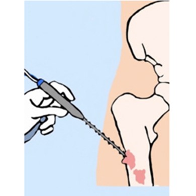
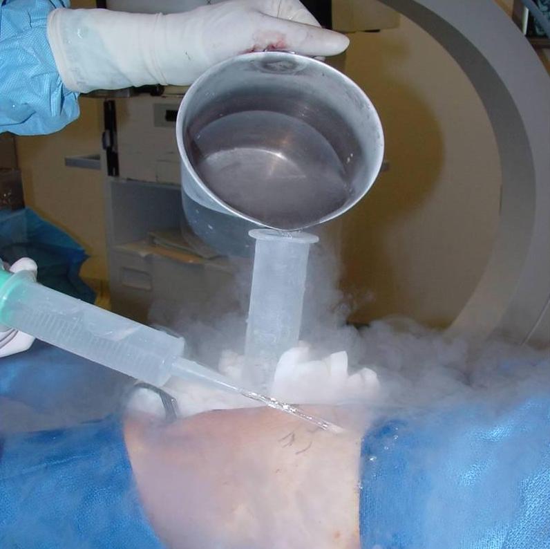
Cryosurgery Video
Dr. James Wittig narrates a video illustrating the surgical technique for resection of a sacrococcygeal chordoma, using cryosurgery as an adjuvant therapy. | WATCH VIDEO