What is an Adamantinoma?
An adamantinoma is a rare, slow-growing bone sarcoma (cancer). It might arise from a pre-existing condition from the bone called an osteofibrous dysplasia. Since these tumors are slow-growing, they rarely spread to other sites in the body.
Who is usually affected?
- • Most common in 20 to 40 year olds.
- • Peak age is in the third decade of life.
- • Males and females are affected equally.
Causes
- • There is a recurrent pattern of numerical abnormalities including extra copies of chromosomes 7, 8, 12, 19 and 21.
- • There is a chromosomal translocation.
- • Many are thought to to arise from osteofibrous dysplasia.
Common Bones Involved
- • Most commonly occurs in the center of the shinbone, which is known as the tibia bone.
- • Occasionally in the jaw, forearm, hands or feet.
- • This includes the fibula, ulna, femur, humerus and radius.
Signs and Symptoms
- • Symptoms include a slow-growing swelling with or without pain.
- • A break due to the tumor weakening the bone.
- • When the tumor develops specifically in the tibia, the lower leg may have a deformed appearance due to curving (bowing) of the bone.
- • Bone deformity and fracture due to the cancer are the main reasons patients seek medical attention.
- • 50% of cases have a history of localized trauma.
Biological Behavior
- • Over 80% of these tumors are at least 5 cm in size.
- • There are two types: Classic vs. Differentiated.
- • Classic type grows beyond the bone cortex, is seen in older patients, and sometimes metastasizes, or spreads to other areas of the body.
- • Differentiated type is confined to the bone cortex, seen in earlier age, and does not metastasize.
- • Sarcomas are categorized into low-grade, intermediate-grade or high-grade lesions based on a variety of biological factors. This grade classification represents their biologic aggressiveness and correlates with the likelihood of the cancer spreading to other parts of the body.
Diagnosis
- • Biopsy: The diagnosis of musculoskeletal tumors is based on clinical evaluation, radiological imaging, and pathological findings from a biopsy. A biopsy is when the surgeon cuts out a piece of the tumor and looks at it under a microscope. This is important for confirmation of the diagnosis for accurate classification before creating a definitive treatment plan. Open incisional biopsy is the most reliable of current available diagnostic methods.
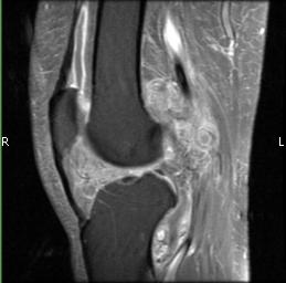
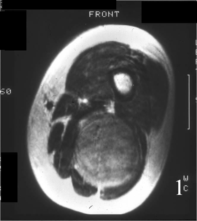
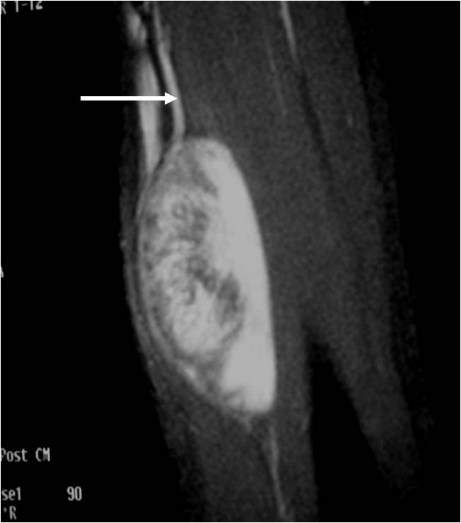
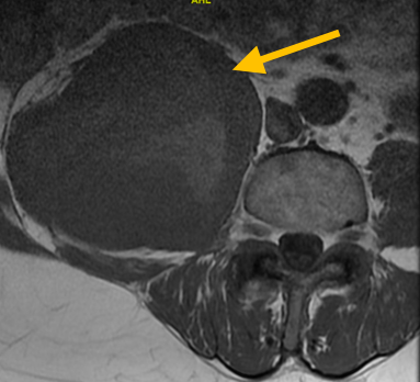
- • Imaging: Staging studies for a sarcoma include plain radiographs (X-rays), computed tomography (CT), and magnetic resonance (MRI) scans. X-Ray remains the key imaging in evaluating a bone lesion. The CT scan allows the doctor to understand the extent of the tumor. The MRI scan provides information on the tumors relation to vital structures, including nerves and blood vessels. Further imaging may be needed including CT of the chest and positron emission tomography (PET) scans to rule out the spread of the cancer to other parts of the body. Sometimes a test called a bone scan is ordered, which is a whole-body method of understanding the entire skeleton.
Risk to your limbs
Adamantinomas are cancerous tumors that, if left unchecked, will grow and destroy your normal bone. As the tumor grows, the bone is weakened and you are at an increased risk of breaking the bone due to the tumor, called a pathological fracture.
Radiographic imaging is used to help form a diagnosis of ABC. These include X-Ray, MRI, CT and Bone Scans.
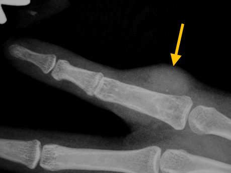
An example of an Adamantinoma X-Ray is shown.
Treatment of Adamantinoma
Surgery

Patients treated with wide excision surgery have a lower rate of the tumor coming back (recurrence). The main treatment of this is surgical removal, wide excision with clear margins. After an en bloc, wide resection, reconstruction is performed. Amputation does not increase survival rate compared to limb saving surgery.
Bone Grafting and Fixation
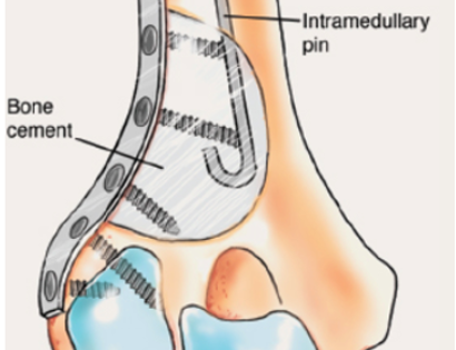
The empty bone cavity is usually filled with bone graft or bone cement. Bone can be donated (allograft) or taken from the patient themselves (autograft). Fixation devices, such as a plate and screws, may be used in specific situations to prevent postoperative fracture
Radiographic Image of an Adamantinoma
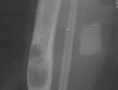
On an X-Ray, also called a radiograph, an adamantinoma can be diagnosed by looking at certain features. Most commonly seen in adamantinoma is a sharply defined osteolytic defect which is lobulated, multicystic, or a “soap bubble”.