What is a Hemangioendothelioma?
A hemangioendothelioma is a cancerous tumor of bone, made of cells named endothelial cells. There is a low rate of metastasis, or spreading, to other parts of the body.
Who is usually affected?
- • This condition is very rare, with only one in one million people diagnosed worldwide.
- • There is a wide distribution of age, ranging from 5-70 years old.
- • The majority of cases occur between 15 to 40 years of age.
- • There is a slight male dominance, with a male to female ratio of 1.5:1.
Causes
- • Most hemangioendotheliomas have a genetic mutation, called a chromosomal translocation. This is when chromosomes break apart and combine together again in the wrong way.
- • The WWTR gene and the CAMTA1 gene are associated genetic markers. The YAP gene and the TFE3 gene are associated genetic markers but less common.
Common Bones Involved
- • Most commonly seen in the skull, spine, ribs and lower extremity.
Signs and Symptoms
- • Pain and swelling.
Biological Behavior
- • There is no heredity component to this condition, so it is not passed down in families.
- • There is a low rate of metastasis, or spreading, to other parts of the body.
- • This cancer is caused by uncontrolled cell growth in the cells that make up the blood vessels, called endothelial cells.
- • A very rare type of cancer.
Diagnosis
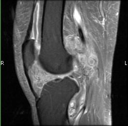
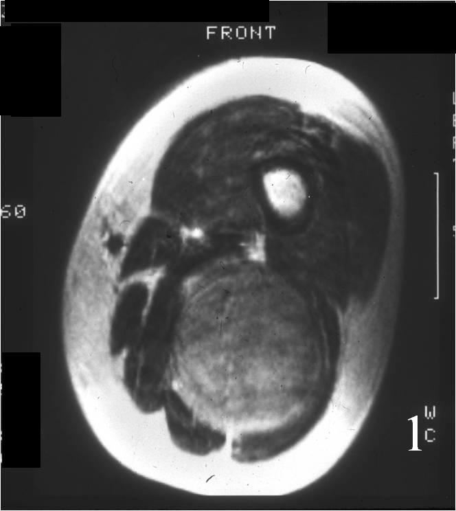
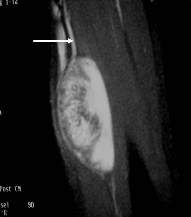
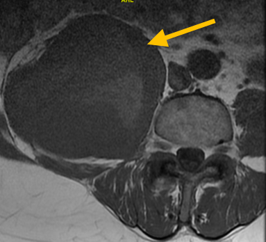
- • The work-up often consists of a physical examination, X-rays, CT scans, MRI, and sometimes bone scans are required.
- • CT of the chest is necessary to check for spread, or metastasis, of the cancer.
- • The diagnosis is often confirmed with a biopsy, which means taking a sample of tumor and having it analyzed under a microscope by a pathologist.
Risk to your limbs
Hemangioendotheliomas are cancerous aggressive tumors that, if left unchecked, will grow and destroy your normal bone. Clinically, local pain and swelling may be the first signs of a growing hemangioendothelioma. As the tumor slowly grows, the bone is weakened and you are at an increased risk of breaking the bone due to the tumor (called a pathological fracture). They may also spread to your lungs or other bones.
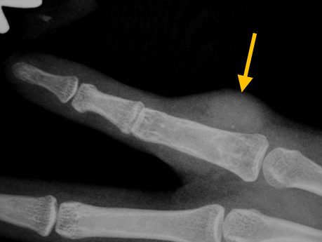
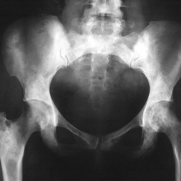
Radiographic imaging is used to help form a diagnosis. These include X-Ray, MRI, CT and Bone Scans.
An example of a X-Ray is shown.
Treatment of Hemangioendothelioma
Limb sparing surgery is the preferred treatment. In low-grade lesions, the standard of treatment is commonly curettage, or scooping out the tumor with a instrument called a curette. This is often sufficient for curing the disease, if the cancer has not spread to other parts of the body (metastasis). Other treatments include surgical ablation and radiation.
Intralesional Curettage
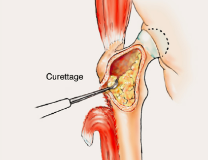
Intralesional Curettage means to scoop the tumor out using a spoon-like tool called a curette. This is a surgery that aims to remove the mass and restore the bone so that the patient can get back to normal function. The hemangioendothelioma is identified within the bone and scooped, or curetted, out. The cavity is then shaved down with a Midas Rex Drill, which is similar to a dental drill. This drill removes more tumor cells.
Surgical Ablation
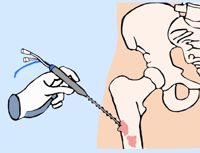
A needle or probe is inserted under CT scan so we can specifically locate the tumor. Once we have located the tumor, radiofrequency waves are aimed and ablate (or melt) the cells.
Radiation

Radiation is a treatment option for some cancers. Radiation therapy is a localized treatment that utilizes high-energy particles or waves to kill cancerous cells. Because radiation therapy is a localized treatment, it only affects the area in which it is set to target and therefore eliminates the risks of damaging healthy cells throughout the body. Not only is it used to treat cancer, but it can also decrease the chances of the cancer from recurring. Lastly, radiation may be used in conjunction with other treatments, such as surgery or chemotherapy, to treat cancers.