What is an Enchondroma?
Bones grow from a cartilaginous growth plate that gradually lengthens and turns into bone as it lengthens. An enchondroma is a benign bone tumor or neoplasm that can be thought of as an island of cartilage within the bone that never turned into normal bone. This type of tumor is usually found incidentally due to injury, as this tumor may weaken the bones and predispose them to fracture.
Who is usually affected?
- • There is a wide distribution of 5-70 years of age.
- • The majority of cases occur between 15 to 40 years of age.
- • Men and women are equally affected.
Causes
- • The exact cause of enchondromas is unknown, Enchondromas are not passed down to your children (non-hereditary).
Common Bones Involved
- • Hands
- • Feet
- • Humerus
- • Femur
Signs and Symptoms
- • Depends on the location.
- • Bones in the fingers may be painful due to stress fractures.
- • Enchondromas found in the long bones (femur, humerus) are found incidentally as no symptoms are usually experienced.
Biological Behavior
- • Enchondromas are benign tumors that do not grow (indolent).
- • There is very minimal risk of the tumor coming back once removed and if recurrence does occur it will be considered cancerous.
- • Rarely, enchondromas can transform into a cancerous (malignant) tumor.
Diagnosis
- • The work-up often consists of a physical examination, X-rays, CT scans, MRI, and sometimes bone scans are required.
- • The diagnosis is often confirmed with a biopsy, which means taking a sample of tumor and having it analyzed under a microscope by a pathologist.
Risk to your limbs
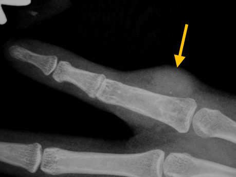
Enchondromas are benign tumors that may weaken the bone primarily in the hands and feet, leading to the bone breaking (called a pathological fracture).
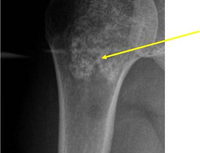
Radiographic imaging is used to help form a diagnosis. These include X-Ray, MRI, CT and Bone Scans.
An example of an enchondroma X-Ray is shown.
Treatment of Enchondroma
If there is a chance of pathological fracture or a pathological fracture already occurred, the surgery used to treat this condition is known as intralesional curettage and bone grafting.
Intralesional Curettage
Intralesional Curettage means to scoop the tumor out using a spoon-like tool called a curette. This is a surgery that aims to remove the mass and restore the bone so that the patient can get back to normal function. The enchondroma is identified within the bone and scooped, or curetted, out. The cavity is then shaved down with a Midas Rex Drill, which is similar to a dental drill. This drill removes more tumor cells.
Bone Grafting and Fixation
The empty bone cavity is usually filled with bone graft or bone cement. Bone can be donated (allograft) or taken from the patient themselves (autograft). Fixation devices, such as a plate and screws, may be used in specific situations to prevent postoperative fracture. This example of an X-ray shows the bone graft filled in nicely. It looks particulate. In this case, the patient was placed in a cast to protect from fracture and allow the graft to heal.
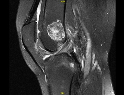
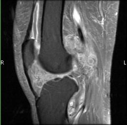
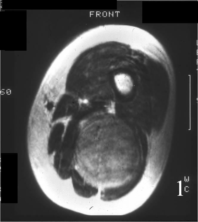
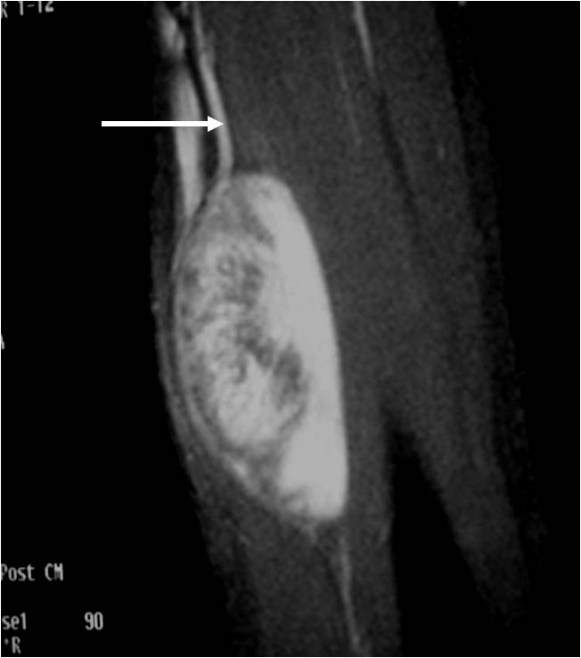
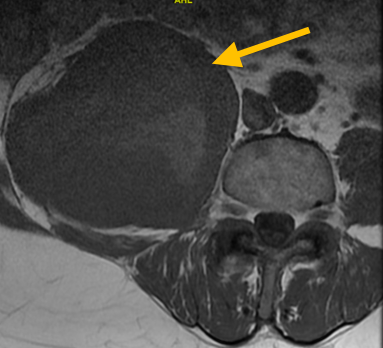
Example of MRI showing an Enchondroma