What is an Epithelioid Hemangioendothelioma?
Who is usually affected?
- • Usually in ages 30-50 years old yet also occurs in younger and older people.
- • Very rare with 1 in 1 million people diagnosed worldwide.
Causes
- • There is thought to be a genetic mutation involved in developing this condition. This is called a chromosomal translocation mutation.
Signs and Symptoms
- • Signs and symptoms include discomfort, pain, nausea, cough, trouble breathing, fever, swelling, weight loss, skin lumps or bumps and broken bones.
Common Sites Involved
- • Most common sites are liver, lungs and bone.
Biological Behavior
- • The biological behavior of EHE is diverse greatly between indolent and aggressively malignant.
Diagnosis
- • This condition is hard to diagnose.
- •The work-up often consists of a physical examination, X-rays, CT scans, MRI, and sometimes bone scans are required. CT scans can be used to check for subtle mineralization that may help with the diagnosis
- • CT of the chest is necessary to check for pulmonary metastases. The lungs and other bones are the to most common sites for the tumor to spread.
- • The diagnosis is often confirmed with a biopsy, which means taking a sample of tumor and having it analyzed under a microscope by a pathologist.
Risk to your limbs
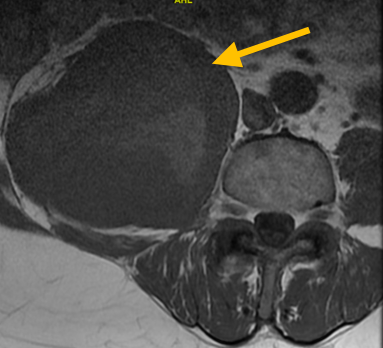
Epithelioid Hemangioendothelioma are cancerous aggressive tumors that, if left unchecked, will grow and destroy your normal bone. As the tumor slowly grows, the bone is weakened and you are at an increased risk of breaking the bone due to the tumor (called a pathological fracture). They may also spread to your lungs or other bones.
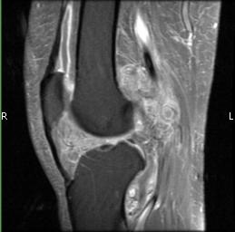
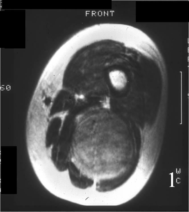
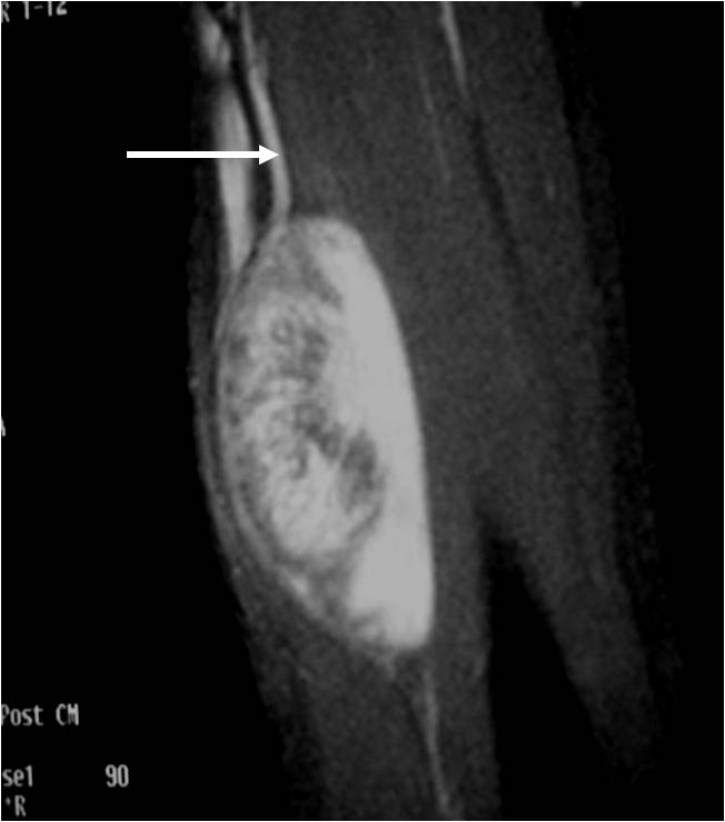
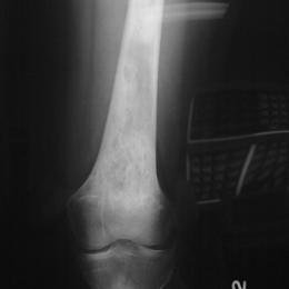
Radiographic imaging is used to help form a diagnosis. These include X-Ray, MRI, CT and Bone Scans.
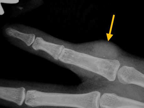
An example of an X-Ray is shown.
Treatment of Epithelioid Hemangioendothelioma
This condition is very rare and thus there is no standard of treatment. Treatment is very individualized depending on where it is in the body and if it has spread. Treatments are surgery, chemotherapy, radiation, targeted therapy, immunotherapy and embolization.
Intralesional Curettage
Intralesional Curettage means to scoop the tumor out using a spoon-like tool called a curette. This is a surgery that aims to remove the mass and restore the bone so that the patient can get back to normal function. The ABC is identified within the bone and scooped, or curetted, out. The cavity is then shaved down with a Midas Rex Drill, which is similar to a dental drill. This drill removes more tumor cells.
Cryosurgery
It is a specialized technique that only a handful of surgeons in the country know how to perform. Once the tumor is removed, liquid nitrogen may be poured into the bone cavity to freeze the area to sub zero temperatures in order to kill microscopic tumor cells. This reduces the chances of the tumor coming back to less than 5%. Warm fluid is also used to prevent normal tissues from freezing.
Bone Grafting and Fixation
The empty bone cavity is usually filled with bone graft or bone cement. Bone can be donated (allograft) or taken from the patient themselves (autograft). Fixation devices, such as a plate and screws, may be used in specific situations to prevent postoperative fracture. This example of an X-ray shows the bone graft filled in nicely. It looks particulate. In this case, the patient was placed in a cast to protect from fracture and allow the graft to heal.