What is an Osteoid Osteoma?
A small and noncancerous bone tumor where the cells in the tumor, called osteoblasts, make new bone. It is almost always very painful and the pain is usually turned off by taking aspirin or NSAIDS. They are very irritating to the bone and result in significant swelling in the bone and surrounding tissues.
Who is usually affected?
- • Most common in the second decade of life, with 75% of patients younger than 25 years old.
- • It is rarely seen in patients over 30 years old.
- • Males are affected 3x more than females.
Causes
- • The cause is unknown.
Common Bones Involved
- • Common in long bones of the lower extremities, including the femur and tibia.
- • Most common in the femur, at the top of the femur.
- • When it occurs in the spine it can cause scoliosis (spinal curvature).
Signs and Symptoms
- • Signs and symptoms include pain that is often worse at night, pain that is relieved by aspirin or NSAIDs, swelling, and sometimes, but not always, a lump.
- • Usually a single lesion that is very painful and is 2 cm in size or smaller.
Biological Behavior
- • A benign (noncancerous) bone tumor where the cells in the tumor called osteoblasts make new bone.
- • It looks identical under a microscope to an osteoblastoma. An osteoblastoma is a rare, benign aggressive bone forming tumor that continues to grow and destroy bone.
- • They are usually anywhere from a few millimeters in size up to 2 cm. The growth of an osteoid osteoma is limited and does not grow larger than 2 cm.
Diagnosis
- • It can be difficult to diagnose especially when it’s small and adjacent to a joint. In some instances, children are diagnosed as having juvenile rheumatoid arthritis because of the extensive swelling in the joint and can go a long time without an accurate diagnosis.
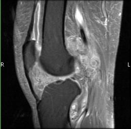
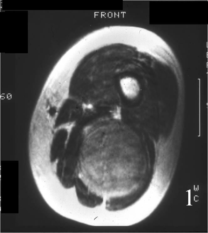
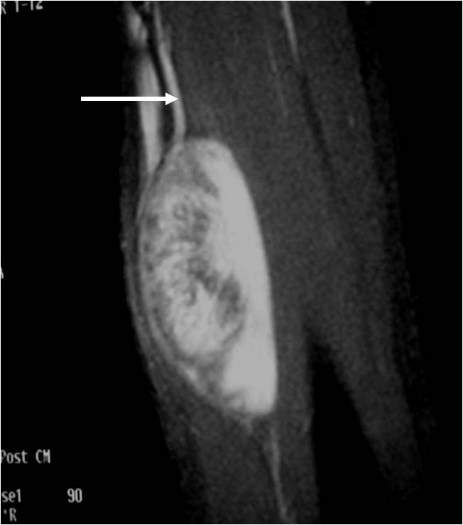
- • Scans include X-ray, CT, MRI, Bone scans. When it is very small and only a few millimeters it can be difficult to see on an Xray and MRI. The MRI usually shows extensive swelling around the tumor that can make it difficult to see the actual tumor on the MRI.
- • The diagnosis is often confirmed with a biopsy, which means taking a sample of tumor and having it analyzed under a microscope by a pathologist.
Risk to your limbs
As the tumor slowly grows, the bone is weakened and you are at an increased risk of breaking the bone due to the tumor (called a pathological fracture). They may also spread to your lungs or other bones.
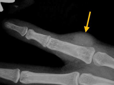
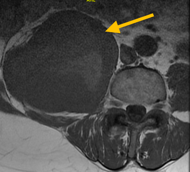
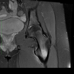
Radiographic imaging is used to help form a diagnosis. These include X-Ray, MRI, CT and Bone Scans.
An example of an osteoid osteoma is shown.
Treatment of Osteoid Osteoma
Treatment is usually with minimally invasive percutaneous CT guided radiofrequency ablation for osteoid osteomas in the long bones, pelvis and sometimes the spine. If not causing any symptoms, it may be recommended that the tumor be left alone.
Radiofrequency Ablation
The tumor is heated and destroyed with a high-frequency electrical current, with minimal damage to the surrounding tissues. Ablation treatment is effective over 90% of the time. Advantages of this treatment include rapid pain relief within 2-3 days after the procedure, no overnight hospital stay, return to normal activities within a week after the procedure and minimal damage to bones and muscles with no structural damage.
“Burr-Down” Resection
The hands, wrist and feet and some other areas may be treated with an open surgical procedure called a “Burr-Down” resection where a high speed drill similar to a dental drill is used to shave the bone and remove the tumor.
NSAIDs
Over the counter medications can be used to relieve pain including aspirin, ibuprofen, naproxen and other NSAIDs. Non-steroidal anti-inflammatory drugs (NSAIDs) are medications commonly used for pain relief and reduce inflammation and fevers.