What is a Parosteal Osteosarcoma?
Who is usually affected?
- • Most common in early adulthood and middle age, 20-40 years old.
- • Affects females more than males. 2:1 sex ratio.
Causes
- • The exact cause of osteosarcoma is not known, but it is believed to be due to DNA mutations inside bone cells.
Common Bones Involved
- • Most common sites include femur, tibia and humerus.
- • 80% of cases occur in the femur and most on the back surface of the femur (thigh bone near the knee).
Signs and Symptoms
- • Signs and symptoms include a painless, slowly growing mass. It usually exists for several years. If the mass is large and on the back of the femur an inability to bend the knee is seen.
Biological Behavior
- • Low risk of spreading (metastasizing). Approximately a 10 percent risk of spreading.
- • Some tumors dedifferentiate and can become more aggressive with a higher risk of spreading to the lungs and other bones. Parosteal osteosarcomas that back invade into the canal of the bone are usually higher grade and have higher risk of spreading.
Diagnosis
- • The work-up often consists of a physical examination, X-ray, MRI, and CT scans.
- • CT of the chest is necessary to check for pulmonary metastases. The lungs and other bones are the to most common sites for the tumor to spread.
- • The diagnosis is often confirmed with a biopsy, which means taking a sample of tumor and having it analyzed under a microscope by a pathologist.
Risk to your limbs
Parosteal Osteosarcomas are cancerous tumors that, if left unchecked, will grow and destroy your normal bone. As the tumor slowly grows, the bone is weakened and you are at an increased risk of breaking the bone due to the tumor (called a pathological fracture). They may also spread to your lungs or other bones.
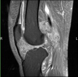
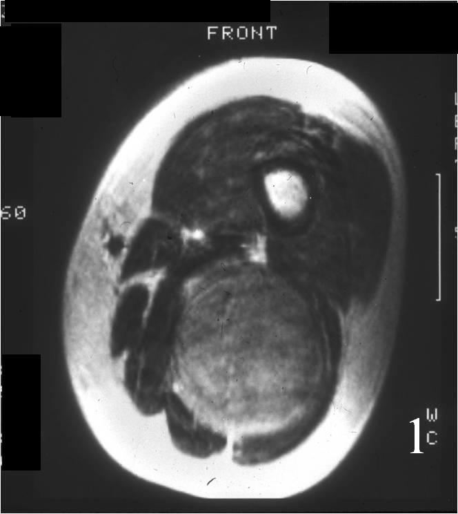
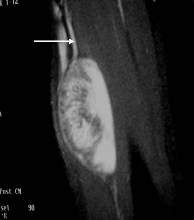
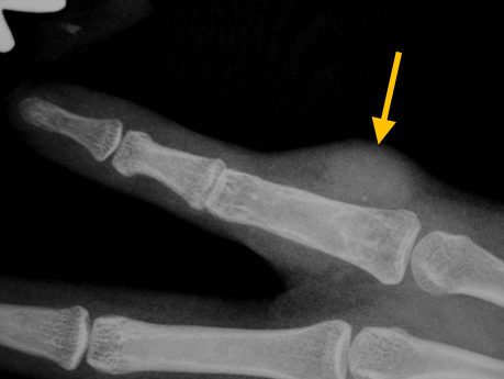
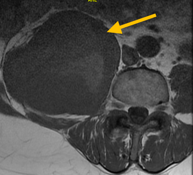
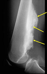
Radiographic imaging is used to help form a diagnosis. These include X-Ray, MRI, CT.
An example of an MRI is shown.
Treatment of Parosteal Osteosarcoma
Most commonly be treated with limb-saving surgical resection, with wide margins and reconstruction with a prosthesis depending on exact location and size without chemotherapy or radiation. Excellent prognosis with 80-95% long term survival (approximately 90% of patients are cured with surgery alone). High grade tumors like those that dedifferentiate may need chemotherapy in addition.
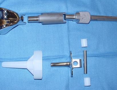
Limb Sparing Surgery
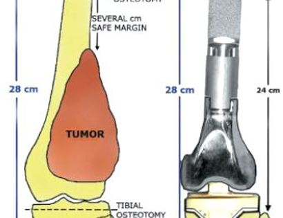
The removal of a tumor from a limb without having to remove the limb itself. The tumor will be removed and samples of tissue will be checked by a pathologist to ensure that only healthy tissue is left after removal. Skeletal reconstruction is done with a metal prosthesis.
Diagram and X-Ray of Prosthesis Inserted
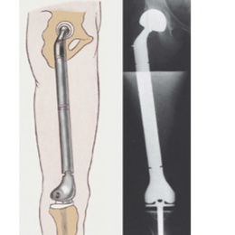
Photo of Metal Prosthesis