What is a Chondroblastoma?
A rare type of noncancerous (benign) bone tumor that begins in is made up of primitive (The type of cells found in an embryo called chondroblasts) cartilage cells. The tumor is benign but also painful, aggressive and destroy bone and sometimes the adjacent joint as it grows. Chondroblastomas grow at the ends of the body’s long bones (called the epiphysis) near the joints. Common bones include the femur, tibia and humerus. They sometimes go undiagnosed and are attributed to sports injuries, growing pains and since adjacent to a joint, forms of arthritis.
Who is usually affected?
- • Typically occurs in children and young adults.
- • 80% of cases are under 25 years of age.
- • More common in males.
- • Males are twice as likely as females to develop the condition.
Signs and Symptoms
- • Pain is the most common symptom.
- • Joint stiffness and joint swelling.
- • Muscle atrophy (wasting away of surrounding muscle from lack of use due to pain).
- • A limp (if tumor is in the lower extremity).
Common Bones Involved
- • Humerus (long bone above your elbow)
- • Femur (long bone above your knee)
- • Tibia (long bone below your knee)
Causes
- There is no known cause of chondroblastomas.
Diagnosis
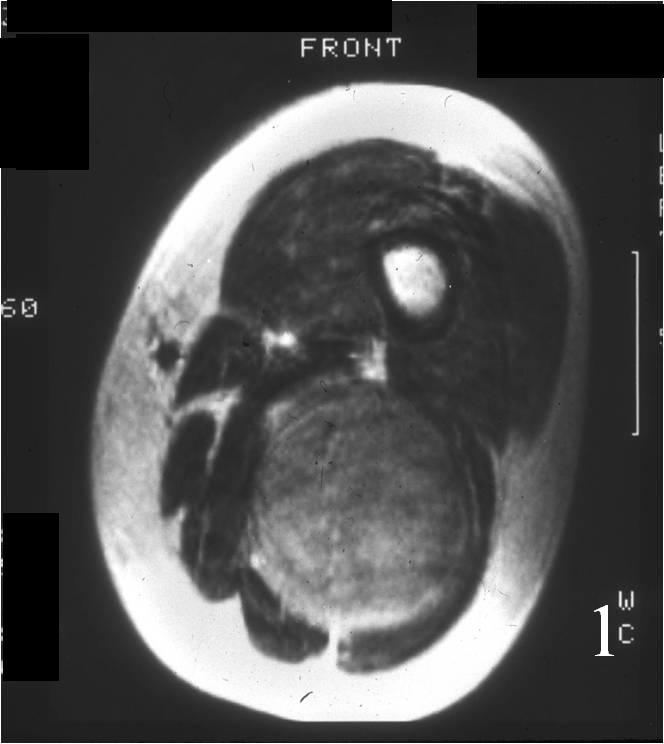
- • The work-up for ABCs often consist of a physical examination, scans including X-Ray, MRI and Bone Scans. A CT scan is sometimes used.
- • The diagnosis is often confirmed with a biopsy, which means taking a sample of tumor and having it analyzed under a microscope. Under a microscope, chondroblastomas look like immature “baby” cartilage cells. Calcifications may be seen that weave through the tumor, resembling a “chicken wire”. Also a “cobblestone” appearance is described by pathologists. In many instances the biopsy can be performed at the same time as the definitive surgery.
Biological Behavior
- • Chondroblastoma is one of a few benign tumors with the potential of extremely rare spread (metastasize) to the lungs. However, it is still considered a benign tumor because this is a rare event and these deposits in the lungs sometimes disappear on their own and rarely if ever lead to problems or death.
- • It is usually small in size, from 1 to 4 cm but has the ability to grow uncontrollably to much larger sizes and be extremely destructive.
Risk to your limbs
If chondroblastomas are left untreated, they can continue to grow and destroy the surrounding bone, causing pain and sometimes a fracture.
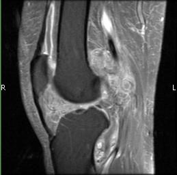
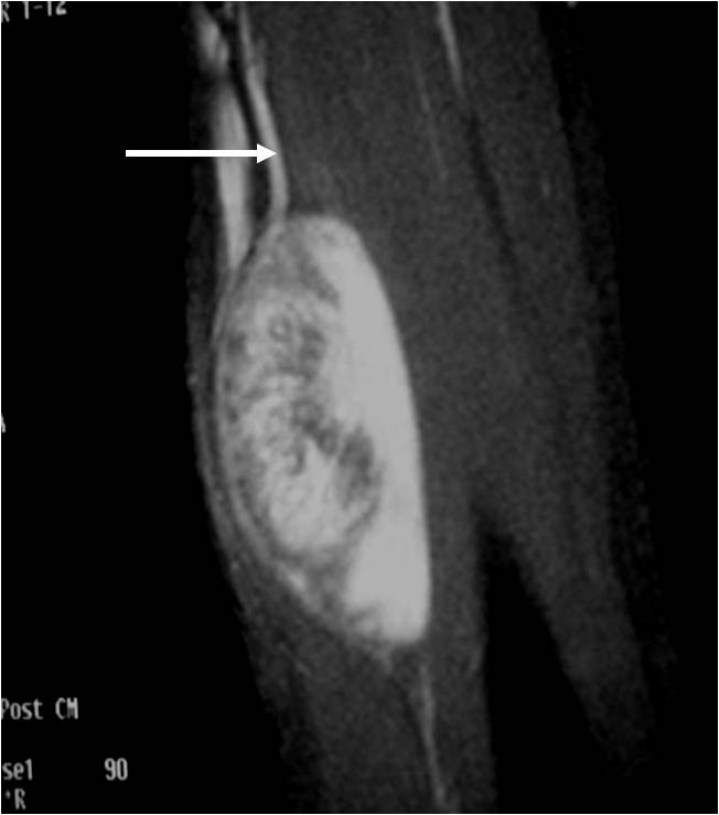
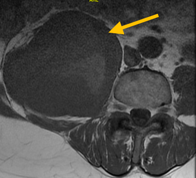
Radiographic imaging is used to help form a diagnosis of Chondroblastoma. Scans include X-Ray, MRI and Bone Scan. CT scan is sometimes used to assess the adjacent bone quality and look for flecks of calcium in the tumor to aid in diagnosis. MRI usually shows extensive swelling around the tumor that is sometimes misinterpreted as a cancer.
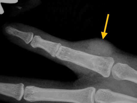
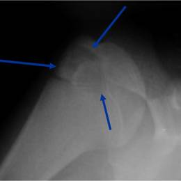
An example of a Chondrosarcoma X-Ray.
Treatment of Chondroblastoma
Treatment most commonly involves surgery to remove the tumor and to prevent damage to the nearby joint. Treatment usually involves curettage, midas rex burring and bone graft. Cryosurgery is often used by Dr. Wittig to kill microscopic cells and reduce the chances of the tumor coming back. Bone cement may also be used to fill the hole in certain instances. Radiofrequency ablation has also been described but is rarely used. Resection may be used when extremely extensive or recurrent. This includes removing an entire section of the bone.
Intralesional Curettage
Intralesional Curettage means to scoop the tumor out using a spoon-like tool called a curette. This is a surgery that aims to remove the mass and restore the bone so that the patient can get back to normal function. The chondroblastoma is identified within the bone and scooped, or curetted, out. The cavity is then shaved down with a Midas Rex Drill, which is similar to a dental drill. This drill removes more tumor cells.
Cryosurgery
It is a specialized technique that only a handful of surgeons in the country know how to perform. Once the tumor is removed, liquid nitrogen may be poured into the bone cavity to freeze the area to sub zero temperatures in order to kill microscopic tumor cells. Warm fluid is also used to prevent normal tissues from freezing. Recurrence can happen in up to 60% of cases. Cryosurgery decreases the risk of it coming back to less than 10%.
Bone Grafting and Fixation
The empty bone cavity is usually filled with bone graft or bone cement. Bone can be donated (allograft) or taken from the patient themselves (autograft). Fixation devices, such as a plate and screws, may be used in specific situations to prevent postoperative fracture.